
Vision Loss Types: Patterns, Severity, and Clinical Findings

 Oct 05, 2026
 10 min read


Overview:
Vision loss can affect what a person sees straight ahead, off to the side, or in specific areas of sight. This article explains how eye doctors describe these patterns, measure their severity, and use examination findings to understand them.
Vision loss is common. In 2024, the Centers for Disease Control and Prevention (CDC) estimated that 32.17 million Americans, or about 9.5% of the population, had presenting visual acuity loss, meaning vision of 20/32 or worse in the better-seeing eye with their usual correction.
That measure, however, does not capture every form of vision loss. One person may see a blurred center, while another can read clearly but miss things off to the side. These differences in location and extent help distinguish one vision loss type from another.
Understanding Vision Loss Beyond the Eye Chart
Different vision problems can sound alike when patients describe them. “Blurry vision,” for example, may refer to a general loss of clarity or to one area of sight that no longer looks right.
Timing can change the meaning of that complaint. A problem that appeared suddenly in one eye raises different questions from vision that has been getting worse over several months.
The examination helps sort that out. An eye chart tells us how clearly someone sees straight ahead, but it cannot account for every visual complaint. What the patient reports needs to be compared with what actually shows up during the examination.
When the two do not fit, that mismatch deserves attention.
Common Vision Loss Types by Pattern
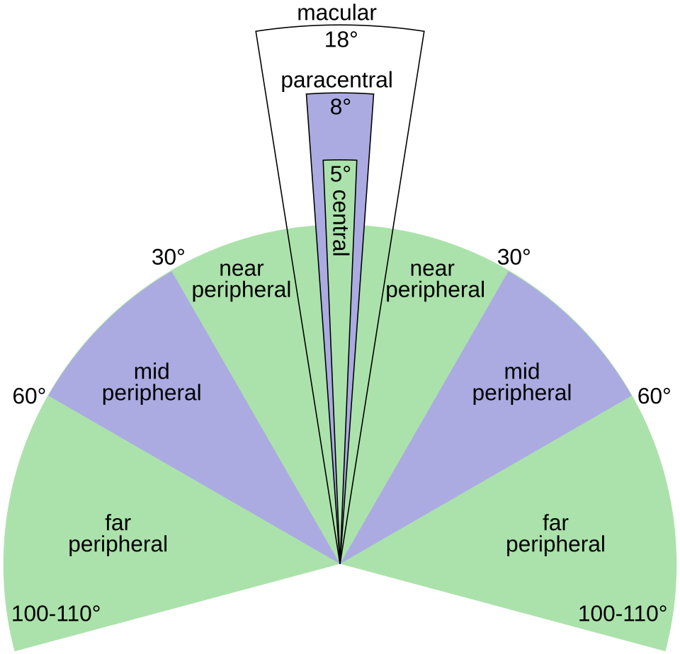
Vision may be affected in the area a person is looking at directly or farther out in the visual field.
The National Eye Institute includes central and peripheral vision loss among the ways low vision can present. Blur, haze, and difficulty seeing in dim conditions may also be part of the experience.
Central and Peripheral Vision Loss
Central vision is used for the detail directly in front of us. Reading a line of text or recognizing a face relies heavily on this part of sight.
The macula, located near the center of the retina, provides much of that fine detail. Damage there can change what a person sees directly ahead. With age-related macular degeneration, for example, the center may look blurred or distorted, and part of an object may seem to be missing.
Peripheral vision refers to what is seen outside that central area. Losing it can be less obvious at first because straight-ahead vision may remain clear. The problem may show up when someone fails to notice a person, vehicle, or object approaching from the side.
Glaucoma can gradually reduce peripheral vision as the optic nerve is damaged. Retinitis pigmentosa can narrow the visual field as well and may make dim conditions harder to navigate.
Scotomas and Visual Field Loss
Central and peripheral vision loss describe where sight is affected, while a scotoma is a localized area of reduced or missing vision within the visual field. The two can overlap: a scotoma may occur in central vision or farther out in the peripheral field.
Some people notice a dark or blurry spot. Others notice the effect instead, such as part of a word disappearing while they read.
Where that missing area appears can matter. A small spot close to the center of vision may interfere with reading or recognizing details. A missing area farther to the side may cause more difficulty with movement and awareness of surroundings.
Larger areas of vision loss can follow recognizable patterns. Hemianopia means that half of the visual field is missing. Quadrantanopia means that about one-quarter is affected.
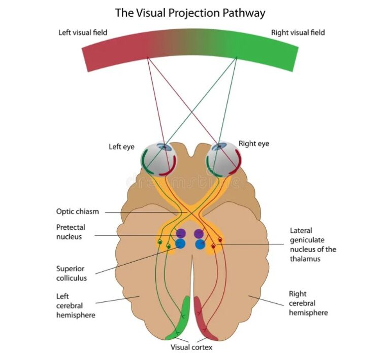
The pattern can give clues about where the problem may be. It could involve the retina, the optic nerve that carries information from the eye, or the nerve pathways that carry visual information toward the brain.
However, I would not base a diagnosis on one visual field test alone. The test needs to be reliable, and sometimes it has to be repeated. I also compare it with the eye examination and what the patient has described.
Blurry, Hazy, and Low-Light Vision
Not every type of vision loss creates a clearly defined missing area. Some patients describe general blur, fogginess, glare, or difficulty seeing after the lights go down.
Many conditions can cause these symptoms. Cataracts, which cloud the eye’s natural lens, can cause haze and glare. A glasses prescription that is no longer correct can cause blur. Some retinal diseases can make night vision worse. Problems involving the clear front surface of the eye, called the cornea, can also reduce clarity.
How the patient describes the problem can provide useful clues. Questions about when it appears and what makes it better or worse can help narrow down the cause. Does vision change with lighting? Does the right prescription make it sharper? Is one eye noticeably worse?
Those answers help me decide what to examine next.

How Vision Loss is Classified by Onset and Severity
Some patients can point to the exact moment their vision changed. Others realize only after several months that they are no longer seeing as well as they used to.
Sudden, Gradual, Temporary, or Persistent Vision Loss
A temporary episode that clears is called transient vision loss. Vision loss that remains is considered persistent. Repeated temporary episodes still deserve attention, even if sight returns between them.
Some sudden visual symptoms need more immediate attention. New flashes of light, a sudden increase in floaters, or a dark curtain or shadow moving across the vision can be signs of retinal detachment, which happens when the light-sensitive tissue at the back of the eye pulls away from its normal position.
These symptoms require urgent eye care.
HVision Loss in One Eye or Both
Whether the problem affects one eye or both also gives useful information.
Loss that truly affects only one eye usually points to a problem in that eye or in the optic nerve connected to it. When both eyes are involved, the cause may affect both eyes or the parts of the nervous system that process vision.
Testing each eye separately helps make that distinction.
Measuring the Severity of Vision Loss
Severity is another part of the assessment. The standard eye chart measures how clearly a person sees fine detail. Doctors can also test how much side vision remains.
Daily function matters as well. Two people with similar test results may not experience the same difficulty because their work, mobility, and daily activities are different.
When vision loss is being assessed for disability, those clinical measurements may also need to be compared with a formal legal definition.
Under U.S. Social Security rules, statutory blindness includes vision of 20/200 or worse in the better eye after the best possible correction. A person may also meet the standard if the remaining field of vision is extremely narrow, measuring 20 degrees or less at its widest point.
This legal definition does not mean that the person has no sight at all. Many people who meet the standard still have some usable vision.
Clinical Findings That Help Explain Vision Loss
After the pattern, timing, and extent of the vision loss are established, the eye examination can show whether the clinical findings match the patient’s symptoms. If they do not, the discrepancy needs a closer look.
Tests Used to Evaluate Vision Loss
The examination usually begins by measuring how clearly each eye sees and whether the correct prescription improves vision.
A visual field test can then show whether certain areas are being missed. Color vision and the pupils’ response to light can also provide useful information when an optic nerve problem is suspected.

A dilated eye examination allows a closer look at the back of the eye, especially the retina and optic nerve. It can also make changes around the macula easier to see.
Sometimes the examination needs to go further. Optical coherence tomography, or OCT, uses light to produce detailed cross-sectional images of the retina and optic nerve. These scans can reveal changes that are difficult to see during a routine examination.
Retinal photographs are useful for comparison over time. If the findings suggest that the problem may come from the visual pathways or the brain rather than the eye itself, imaging such as a CT scan or MRI may be considered.
Test results also have to be reliable. A visual field test, for example, can be affected if the patient has trouble keeping their eyes in position or gives inconsistent responses. When that happens, I may repeat the test before deciding that the vision loss is permanent.
Documenting Vision Loss in Medico-Legal Cases
In a medico-legal review, the question is often not simply whether vision loss exists. The record may also need to show when it began and whether the findings support the limitation being reported.
Earlier examinations can be especially useful when they show what the person’s vision was like before ocular trauma or disputed events. Later records can then be compared with that baseline.
When I review whether an event may have caused or worsened the loss, I compare what was documented before the event with what appeared afterward. The question is whether the change in vision is medically consistent with the event being described.
Conclusion: Understanding the Full Pattern of Vision Loss
Vision loss is best understood in context. A symptom, test result, or diagnosis means more when it is considered alongside the rest of the clinical picture.
That becomes especially important when vision is being tracked over time or reviewed for disability or medico-legal purposes. The record should show a consistent relationship between what the patient reports and what the examination supports.
Frequently Asked Questions
Can You Have Vision Loss Even If You Read an Eye Chart Well?
Yes. Reading the letters on a chart mainly tells us about central clarity. It does not show everything a person sees around that central point. Someone may perform well on the chart and still miss part of the visual field.
When Should a Change in Vision Be Treated as Urgent?
A sudden change can be a reason to seek eye care quickly. New flashes or many new floaters are examples. A dark shadow or curtain moving across the vision is particularly concerning because retinal detachment can cause this symptom.
Why Are the Eyes Tested One at a Time?
Testing the eyes separately can reveal a difference that would be harder to notice with both eyes open. If only one eye is affected, the examination can focus more closely on that eye and its optic nerve. A problem involving both eyes leads to a different set of possibilities.

Related Articles
Understanding Vision Impairment After Ocular Trauma
Learn how ocular trauma is classified, diagnosed, and litigated. Explore why expert ophthalmic interpretation is critical to accurately valuing vision loss claims.
 August 28, 2026
 9 min read


When Vision Loss Is Alleged: How Ophthalmology Experts Assess Medical Causation
Learn how Ophthalmology Experts Assess determine vision loss, identify the underlying condition, and analyze what may have caused it.